
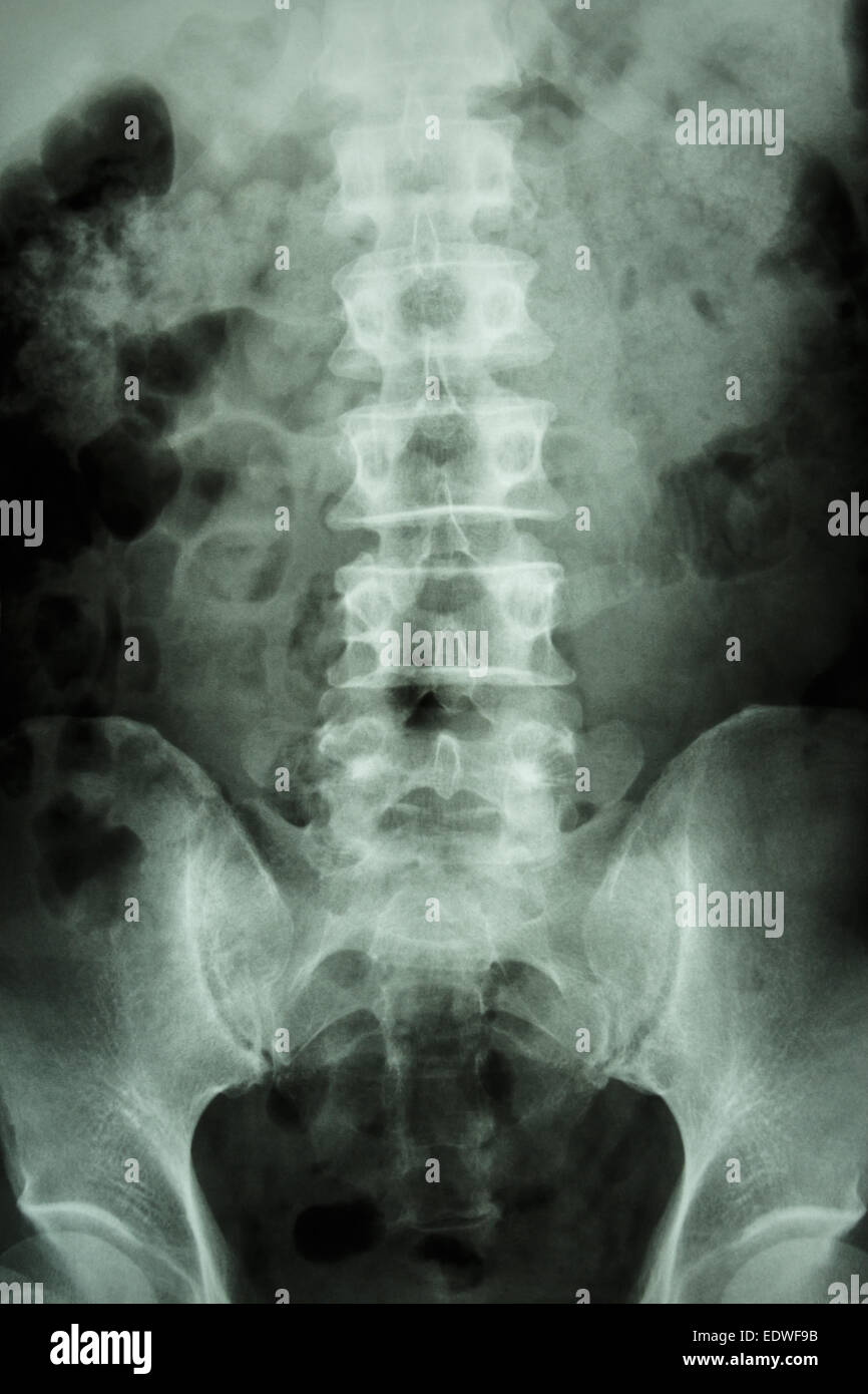
Sacrum Xray
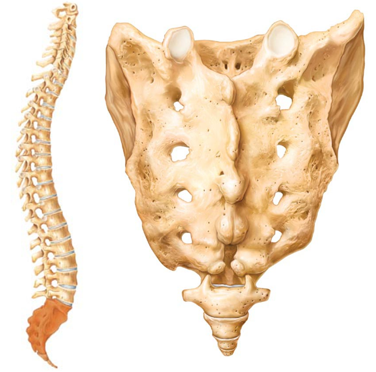
The sacrum has 5 small, fused vertebrae. The 4 coccygeal vertebrae fuse to form 1 bone, called the coccyx or tailbone.. X-rays of the spine, neck, or back may be performed to diagnose the cause of back or neck pain, fractures or broken bones, arthritis, spondylolisthesis (the dislocation or slipping of 1 vertebrae over the 1 below it.

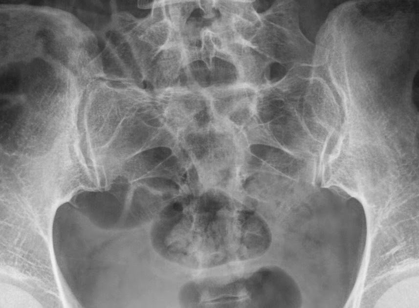
Normal sacrum Xrays Image
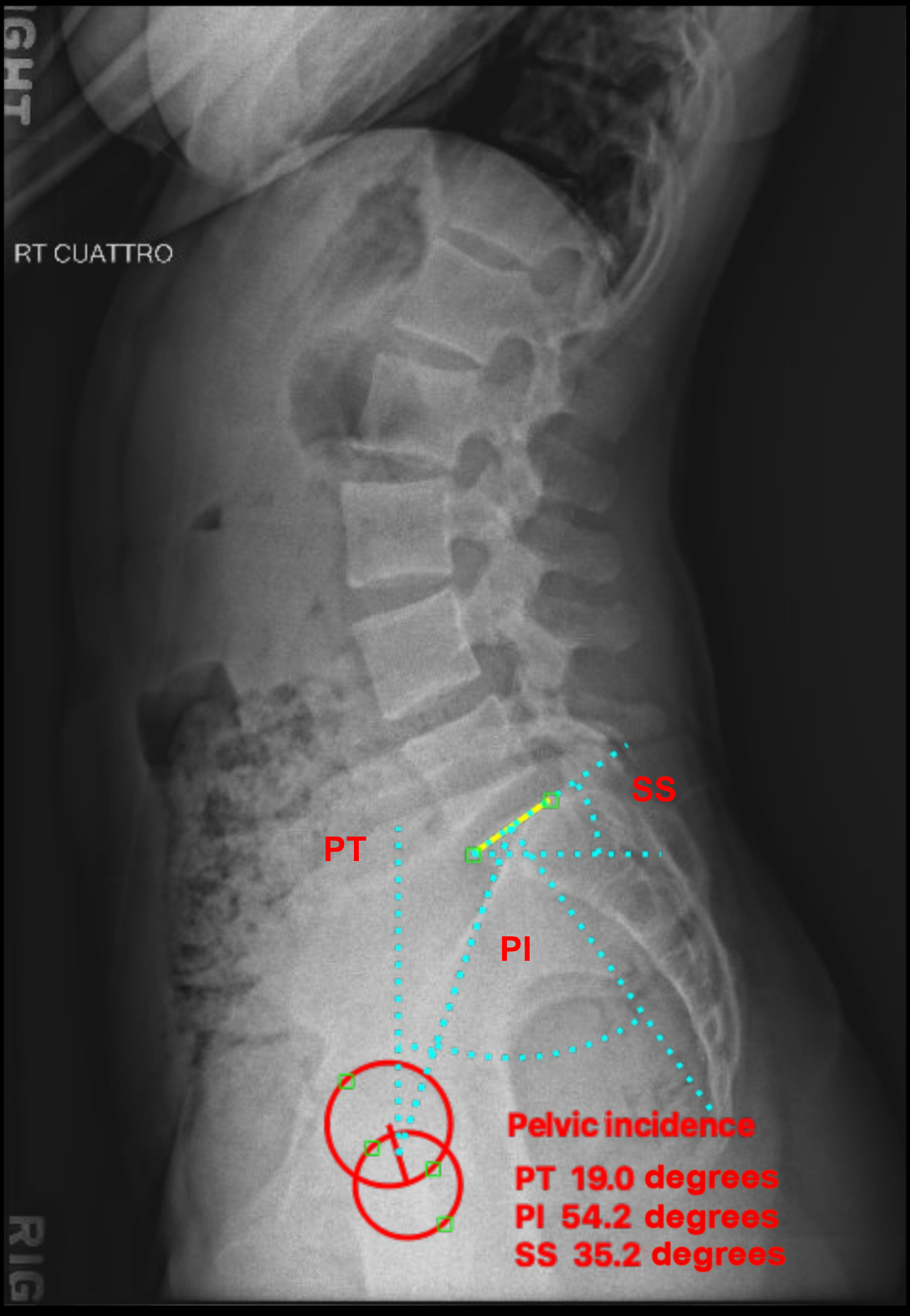
X-ray examination of the sacrum demonstrate fracture and pathology in sacral, sacrum should be foreshortened in AP projection, the Sacroilliac joint and the L5 to S1 joints.
Fractured Sacrum Fracture Treatment
Commonly, coccydynia (coccygodynia) occurs after trauma and appears with normal imaging features at static neutral radiography, but dynamic imaging with standing and seated lateral radiography may reveal pathologic coccygeal motion that is predictive of pain.
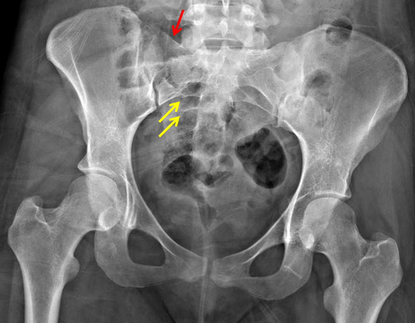
right sacral ala fracture sacral ala fx zones and management Shotgnod
The bony pelvis consists of the sacrum, coccyx and a pair of innominate bones. The innominate bones are formed from three separate bones - the pubis, ilium and ischium. In childhood, they are joined by the triradiate cartilage that unites at puberty to compose the acetabulum, which articulates with the proximal femur at the hip joint.
Cureus Osteitis Condensans Ilii An Cause of Back Pain
The sacrum refers to the bony structure located at the base of the lumbar vertebrae. It forms the posterior pelvic wall(1). The sacrum helps strengthen and stabilize the pelvis(2). Women have a shorter sacrum than men(3). The female sacrum is distributed more obliquely backward, increasing the size of the pelvic cavity.

Pin on Xrays
Technical factors anteroposterior projection centering point central ray midline 5 cm below the level of the anterior superior iliac spine (4 cm above the symphysis pubis) central ray with cephalad angle of 30° (male) to 35° (female) collimation laterally to include both sacroiliac joints superiorly and inferiorly to include the entire sacrum

Imaging Coccygeal Trauma and Coccydynia RadioGraphics
Sacral fractures are common pelvic ring injuries that are under-diagnosed and often associated with neurologic compromise. Diagnosis can made with pelvis radiographs but frequently require pelvic CT scan for full characterization. Treatment may be nonoperative or operative depending on fracture displacement, associated pelvic ring instability.
Sacrum Anatomy Xray
The sacrum is the penultimate segment of the vertebral column and also forms the posterior part of the bony pelvis. It transmits the total body weight between the lower appendicular skeleton and the axial skeleton. Gross anatomy
film xray LS spine (lumbarsacrum) show normal human lumbarsacrum spine Stock Photo Alamy
central ray angled 15° cephalic 3 this angle allows the CR to be perpendicular to the sacrum as it is curved with an anterior concavity and convex posteriorly collimation must adhere to the ALARA principle given the region exposed via the primary beam superior to include the L5/S1 articulation
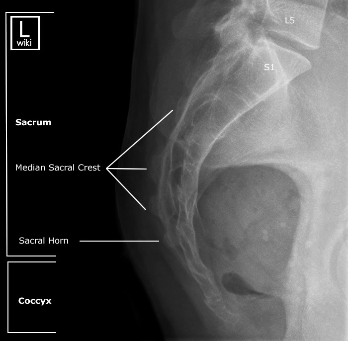
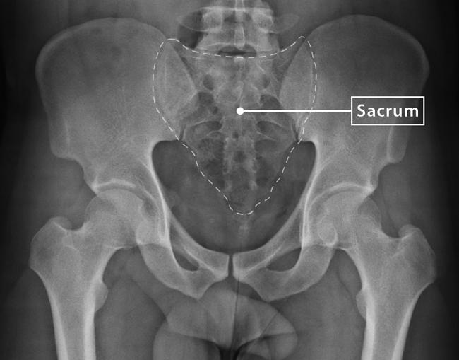
Sacrum Radiographic Anatomy wikiRadiography
Edit article Citation, DOI, disclosures and article data There are several classification systems for sacral fractures, but the most commonly employed are the Denis classification and subclassification systems, and the Isler classification system.

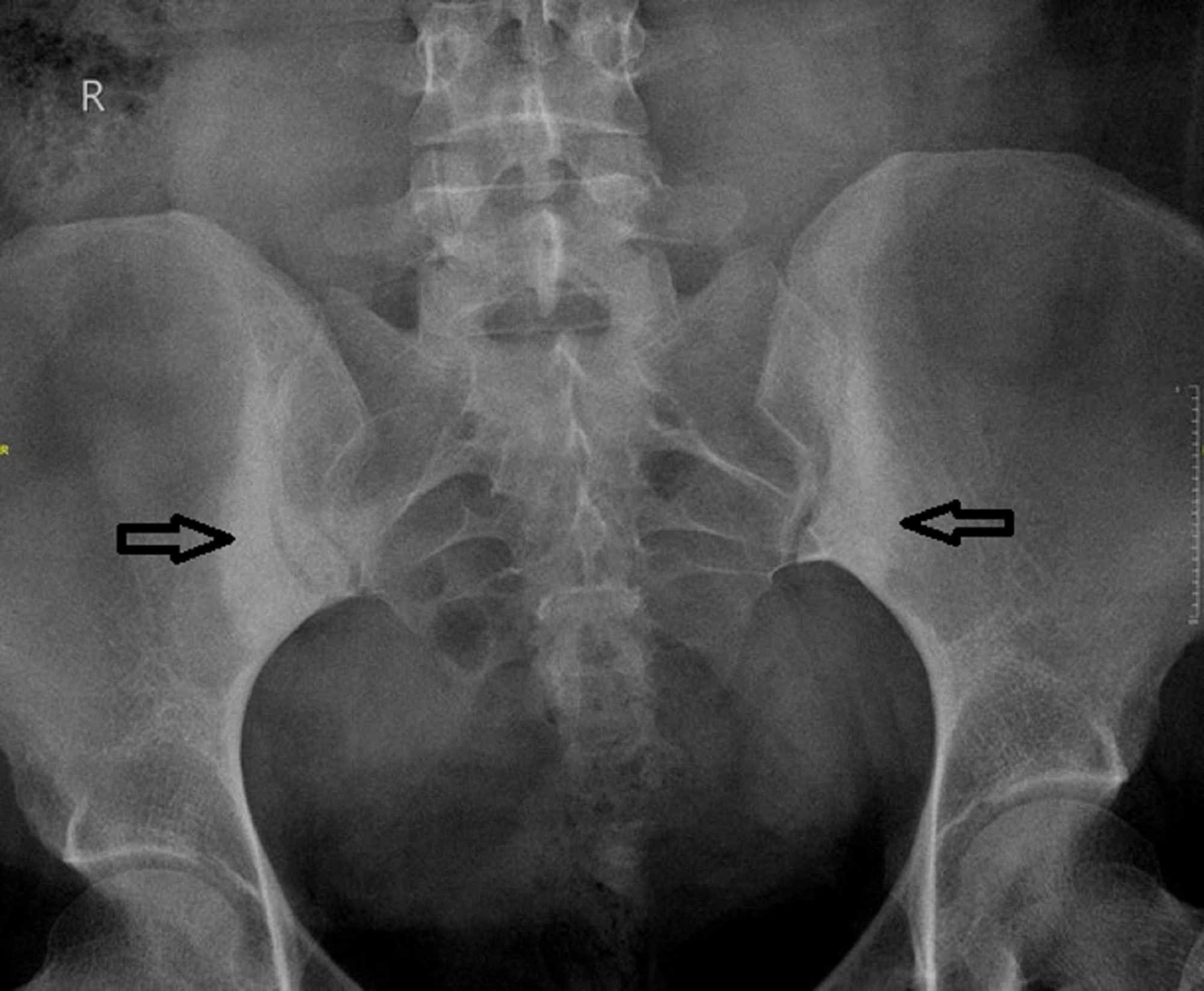
Sacroiliac Joint Dysfunction X Ray
SACRUM ROUTINE VIEWS: AP Lateral AP Sacrum 1. 10 x 12 film 2. Patient supine 3. Bucky 4. Center halfway crest and pubis and midline 5. 40" SID 6. Tube angled 15o cephalad 7. Expiration Lateral 1. 10 x 12 film 2. Bucky 3. Center ASIS and 3" posterior to midaxillary 4. 40" SID 5. Expiration 6. Use lead glove behind patient to absorb scatter

Pin on Radiology
Abstract The sacrum is a structure that is imaged by both general and subspecialty radiologists. A wide variety of disease processes can involve the sacrum either focally or as part of a systemic process. Plain radiographs, although limited in evaluation of the sacrum, should be carefully examined when abnormalities of the sacrum are suspected.
Lumbosacral Spine Plain Radiographs Radiology Key
The sacrum is a triangular-shaped structure composed of five fused vertebral bodies (S1-S5). The sacrum has a critical role in stabilizing the posterior portion of the pelvic ring. At its wider superior aspect, the sacrum forms the lumbosacral joint with the fifth lumbar vertebra above it.

Sacrum Radiographic Anatomy Diagnostic imaging, Radiology imaging, Medical anatomy
Sacrum/Coccyx X-ray Guideline. Routine: 3 views • AP SACRUM with central ray angled 15 degrees cephalad • AP COCCYX with central ray angled 10 degrees caudad • LATERAL Sacrum/Coccyx . Reviewed 2016 AMR . SACRUM COCCYX LATERAL . Author: Alison M. Robinson Created Date:
STOCK IMAGE, xray frontal view of a normal pelvis showing the sacrum and coccyx c athenais
The sacrum and coccyx lateral view is utilized to demonstrate the most distal region of the spine in a lateral position. Indications This projection is commonly used in conjunction with the AP projection or can be used as a sole projection, depending on department protocols.
Your Sacrum & Your Coccyx Clearview Chiropractic
Spondylolisthesis is the anterior displacement of a vertebra relative to the vertebrae below, generally caused by a morphologic abnormality in the pars interarticularis of the vertebral arch. Usually this consists of a defect in the isthmus of the vertebral arch between the superior and inferior articular processes (interarticular spondylolysis).